View Featured Offers >>

Erk Signaling

The MAPK/Erk signaling cascade is activated by a wide variety of receptors involved in growth and differentiation including receptor tyrosine kinases (RTKs), integrins, and ion channels. GPCRs (G-Protein Coupled Receptors) activate the MAPK cascade using a different set of adaptors.

The specific components of the cascade vary greatly among different stimuli, but the architecture of the pathway is similar:

  • Adaptors (Shc, GRB2, Crk, etc.) link the receptor to a guanine nucleotide exchange factor (GEF), i.e. SOS and C3G.
  • The GEF proteins transduce the signal to small GTP binding proteins (Ras, Rap1).
  • The GTP binding protein activates the core unit of the cascade composed of a MAPKKK (Raf), a MAPKK (MEK1/2), and MAPK (Erk). Activated Erk can regulate targets in the cytosol and also translocate to the nucleus where it phosphorylates a variety of transcription factors regulating gene expression.

Activation of Erk (extracellular signal regulated kinase) requires the phosphorylation of Thr202 and Tyr204 amino acid residues.

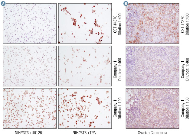
NIH/3T3 cell pellets (Figure A) treated with either TPA (to induce Erk phosphorylation) or U0126 (to inhibit Erk phosphorylation) (Figure B) paraffin embedded ovarian cancer sections.
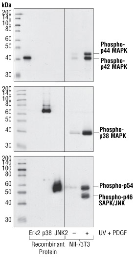
IHC Comparison
  • CST #4370 antibody shows strong cell staining in the TPA treated cells but not in the U0126 treated cells as expected.
  • At a 1:400 dilution, Company 1 antibody shows weak staining in TPA induced cells. At a lower dilution/higher concentration, Company 1 antibody shows strong non-specific staining in the U0126 treated cells.
  • CST #4370 stains ovarian cancer tissues more strongly than the company 1 antibody at the same dilution. When used at a lower dilution/higher concentration, company 1 antibody showed weaker yet comparable staining to CST #4370.
  • Given the background staining by Company 1 antibody in the NIH/3T3 cell pellets treated with U0126, the ovarian cancer tissue staining specificity is questionable.

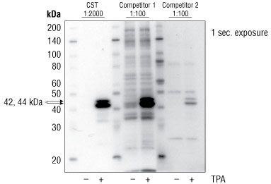
Function: Activation of Erk (extracellular signal regulated kinase) requires the phosphorylation of Thr202 and Tyr204 amino acid residues.

Samples: Jurkat cells were treated with TPA, a phorbol ester that activates PKC to induce Erk phosphorylation.

Western Blot Comparison
  • CST Phospho-p44/42 MAPK (Erk1/2) (Thr202/Tyr204) (D13.14.4E) XP® Rabbit mAb #4370 recognizes the expected double bands at 44 and 42 kDa that correspond to Erk1 and Erk2 respectively.
  • Company 1 phospho-p44/42 MAPK (Erk1/2) (Thr202/Tyr204) mouse monoclonal antibody recognizes the expected double band at 44 and 42 kDa but also shows high background and non-specific bands.
  • Company 2 phospho-p44/42 MAPK (Erk1/2) (Thr202/Tyr204) rabbit polyclonal antibody weakly detects the expected double band at 44 and 42 kDa. TPA strongly induces Erk phosphorylation, therefore the weak signal suggests low antibody binding.
CST #4370 Competitor 1 Competitor 2 Product 4370 Dilution 1:2000 1:100 1:100 Conc. (μg/mL) 0.222 1 10