The healthy immune system employs a series of checkpoints in order to maintain self-tolerance or prevent collateral tissue damage during an immune response. Activation of CD8+ cytotoxic or CD4+ helper T cells occurs through the interaction of the T cell receptors (TCRs) with antigens on the surface of antigen-presenting cells (APCs) in the form of peptide antigen bound to major histocompatibility complex (MHC) molecules (1). In addition, binding of co-stimulatory signaling molecules on T cells (e.g., CD28, ICOS, GITR) with their receptors on APCs (e.g., CD80/CD86, ICOSL, GITRL) can also contribute to T cell activation (1). However, under certain circumstances, T cell receptor engagement is coupled with inhibitory signals that suppress T cell activation and response. These signals are generated by proteins involved in immune checkpoint control such as PD-1, CTLA-4, TIM-3, and LAG3 (2). PD-1 and CTLA-4 immune checkpoint proteins are commonly upregulated in tumor infiltrating T cells, bind their corresponding ligands, PD-L1 (B7-H1) / PD-L2 (B7-DC) and CD80/86 respectively, and downregulate the T cell response. Immune checkpoint ligands are often upregulated in cancer cells as a means to evade immune detection (3-5). Therefore, activating antitumor immunity by blocking immune checkpoint proteins has become the subject of intense research and drug development efforts toward cancer treatment (e.g., Anti-PD-1 immunotherapy) (6).
Below is a table of stimulatory and inhibitory receptor-ligand complexes, which mediate activation or dampening of the T cell response, respectively. Click on linked protein names for additional information and related product lists:
| Cellular Response | T cell | Antigen presenting cell |
|---|---|---|
| Co-stimulatory | CD28 | B7-1 (CD80) or B7-2 (CD86) |
| CD40L | CD40 | |
| TLT-2?** | B7-H3 | |
| OX40 (CD134) | OX40L | |
| 4-1BB (CD137) | 4-1BBL | |
| ICOS | ICOSL | |
| GITR | GITRL | |
| Co-inhibitory | CTLA-4 | B7-1 (CD80) or B7-2 (CD86) |
| PD-1 | B7-H1 (PD-L1) or B7-DC (PD-L2) | |
| Unknown | B7-H3 | |
| Unknown | B7-H4 | |
| Unknown | VISTA | |
| VISTA | Unknown | |
| LAG3 | MHC-Class II | |
| TIM-3 | Galectin-9 |
**Myeloid cell-like transcript 2 (TLT-2) has been shown to express the putative receptor for B7-H3 (7,8). However its co-stimulatory nature and whether it, indeed, interacts with B7-H3 have been challenged (9). Hence the validity of this receptor-ligand interaction is a matter of debate.
There is also increasing interest in understanding the role of immunometabolism through potential therapeutic targets such as IDO and Arginase-1. IDO is an immunosuppressive enzyme involved in tryptophan degradation that is upregulated in many tumors. Arginase-1 is a ubiquitous enzyme highly expressed by myeloid derived suppressor cells (MDSCs) that are often recruited to the tumor microenvironment. In this context, Arginase-1 catalyzes the conversion of L-arginine to urea, thereby depriving T cells of nutrients that are essential for their activation and differentiation (10).
To understand the complex tissue microenvironment under both physiological and pathological conditions such as cancer, profiling immune checkpoint proteins and phenotypic markers is important. This can be achieved by using multiplexed assays for investigating multiple cancer immunotherapy targets and predictive biomarkers in limited and valuable patient samples. Below is a table of immune cell phenotyping markers. Click on linked protein names for additional information and related product lists:
| Phenotypic & Functional Markers | |
|---|---|
| CD3ε | CD4 |
| CD5 | CD8α |
| CD11c | CD14 |
| CD16 | CD19 |
| CD31 | CD36 |
| CD44 | CD45 |
| CD45RO | CD56 (NCAM) |
| CD68 | CD79A |
| CD79B | CD163 |
| CD206 | T-BET |
| F4/80 | FOXP3 |
| Arginase-1 | IDO |
| GATA-3 | Granzyme B |
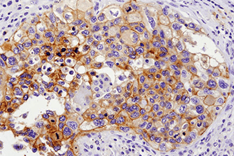
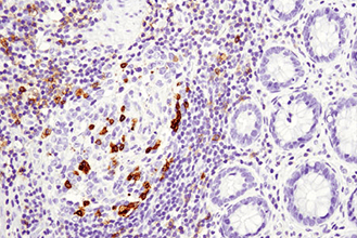
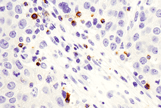
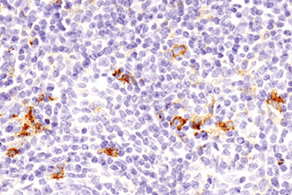
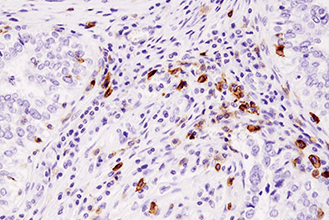
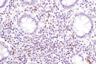
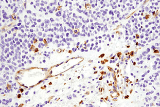
Fluorescent multiplex immunohistochemistry (mIHC), enabling the detection of 6 or more proteins and biomarkers in formalin-fixed, paraffin-embedded (FFPE) tissue samples, is a valuable tool to study immuno-oncology. In mIHC as well as in single/dual-plex chromogenic IHC approaches, using validated antibodies against relevant targets is crucial to obtain reliable results. CST offers human-reactive and mouse-reactive IHC validated antibodies that enable investigators to get more information about biomarker expression, localization, interaction and disease context.