View Featured Offers >>
35347
PD-1 (Intracellular Domain) (D4W2J) & CO-0008-594 SignalStar™ Oligo-Antibody Pair
SignalStar™ Multiplex IHC Kits & Reagents
Oligo Antibody Pair

PD-1 (Intracellular Domain) (D4W2J) & CO-0008-594 SignalStar Oligo-Antibody Pair #35347

SignalStar multiplex immunohistochemical analysis of paraffin-embedded human gastric adenocarcinoma using PD-1 (Intracellular Domain) (D4W2J) & CO-0008-594 SignalStar Oligo-Antibody Pair #35347 (yellow), Pan-Keratin (C11) & CO-0003-488 SignalStar Oligo-Antibody Pair #63566 (red), PD-L1 (E1L3N®) & CO-0005-488 SignalStar Oligo-Antibody Pair #85646 (green), Granzyme B (D6E9W) & CO-0009-594 SignalStar Oligo-Antibody Pair #15194 (orange), TIGIT (E5Y1W) & CO-0002-647 SignalStar Oligo-Antibody Pair #18288 (cyan), CD20 (E7B7T) & CO-0011-647 SignalStar Oligo-Antibody Pair #36775 (pink), CD8α (D8A8Y) & CO-0004-750 SignalStar Oligo-Antibody Pair #62750 (white), CD3ε (D7A6E) & CO-0001-750 SignalStar Oligo-Antibody Pair #51754 (magenta), and ProLong Gold Antifade Reagent with DAPI #8961 (blue). All fluorophores have been assigned a pseudocolor, as indicated. Staining was performed on the BOND RX by Leica Biosystems.
SignalStar immunohistochemical analysis of paraffin-embedded human gastric adenocarcinoma using PD-1 (Intracellular Domain) (D4W2J) & CO-0008-488 SignalStar Oligo-Antibody Pair #17942 (green). All fluorophores have been assigned a pseudocolor, as indicated. Staining was performed on the BOND RX by Leica Biosystems.
SignalStar immunohistochemical analysis of paraffin-embedded human tonsil using PD-1 (Intracellular Domain) (D4W2J) & CO-0008-594 SignalStar Oligo-Antibody Pair #35347 (yellow). All fluorophores have been assigned a pseudocolor, as indicated. Staining was performed on the BOND RX by Leica Biosystems.
SignalStar immunohistochemical analysis of paraffin-embedded human non-small cell lung carcinoma using PD-1 (Intracellular Domain) (D4W2J) & CO-0008-647 SignalStar Oligo-Antibody Pair #56837 (red). All fluorophores have been assigned a pseudocolor, as indicated. Staining was performed on the BOND RX by Leica Biosystems.
SignalStar multiplex immunohistochemical analysis of paraffin-embedded human non-small cell lung carcinoma using PD-1 (Intracellular Domain) (D4W2J) & CO-0008-647 SignalStar Oligo-Antibody Pair #56837 (left, red) and ProLong Gold Antifade Reagent with DAPI #8961 (left, blue) compared to chromogenic immunohistochemical analysis of a serial section of paraffin-embedded human non-small cell lung carcinoma using PD-1 (Intracellular Domain) (D4W2J) XP® Rabbit mAb #86163 (right). All fluorophores have been assigned a pseudocolor, as indicated. Staining was performed on the BOND RX by Leica Biosystems.
SignalStar multiplex immunohistochemical analysis of paraffin-embedded human gastric adenocarcinoma using PD-1 (Intracellular Domain) (D4W2J) & CO-0008-488 SignalStar Oligo-Antibody Pair #17942 (green), TIM-3 (D5D5R) & CO-0010-594 SignalStar Oligo-Antibody Pair #55508 (yellow), LAG3 (D2G4O) & CO-0026-647 SignalStar Oligo-Antibody Pair #40966 (red), and CD3ε (D7A6E) & CO-0001-750 SignalStar Oligo-Antibody Pair #51754 (cyan). All fluorophores have been assigned a pseudocolor, as indicated. Staining was performed on the BOND RX by Leica Biosystems.
SignalStar multiplex immunohistochemical analysis of paraffin-embedded human gastric adenocarcinoma using PD-1 (Intracellular Domain) (D4W2J) & CO-0008-488 SignalStar Oligo-Antibody Pair #17942 (green) and PD-L1 (E1L3N®) & CO-0005-647 SignalStar Oligo-Antibody Pair #52085 (red). All fluorophores have been assigned a pseudocolor, as indicated.

Product Description

SignalStar multiplex immunohistochemistry (IHC) is an advanced technology for labeling multiple proteins simultaneously in tissue samples using specific primary antibodies and fluorescent detection reagents. This technology offers accuracy and reliability in visualizing and analyzing protein expression while maintaining spatial context and tissue architecture.

SignalStar Oligo-Antibody Pairs are compatible with the SignalStar Multiplex IHC Buffer Kits for use in fluorescent multiplex imaging experiments. This product includes the oligo-conjugated antibodies and complementary oligos required for labeling your target protein on up to 10 slides. SignalStar Multiplex IHC Buffer Kits are required to amplify and image the target signal. Multiple oligo-antibody pairs can be conveniently combined into a multiplex panel using the SignalStar Multiplex IHC Panel Builder. SignalStar Multiplex IHC Kits & Reagents are not compatible with all of Cell Signaling Technology® products and protocols that are recommended for use in immunohistochemical assays.

Product Usage Information

Application Dilution
SignalStar™ Leica Bond 1:50 - 1:200
SignalStar™ Manual 1:50 - 1:200

Storage

SignalStar conjugates are supplied in PBS (pH 7.2), less than 0.1% sodium azide, 2 mM EDTA, 0.05% Triton X-100, 2 mg/mL BSA, and 50% glycerol. Complementary oligos are supplied in nuclease-free water. Store at -20°C. Do not aliquot the antibody. All components in this kit are stable for at least 12 months when stored at the recommended temperature.

Specificity / Sensitivity

PD-1 (Intracellular Domain) (D4W2J) XP® Rabbit mAb (SignalStar Conjugate 0008) recognizes endogenous levels of total PD-1 protein.

Species Reactivity:

Human

Source / Purification

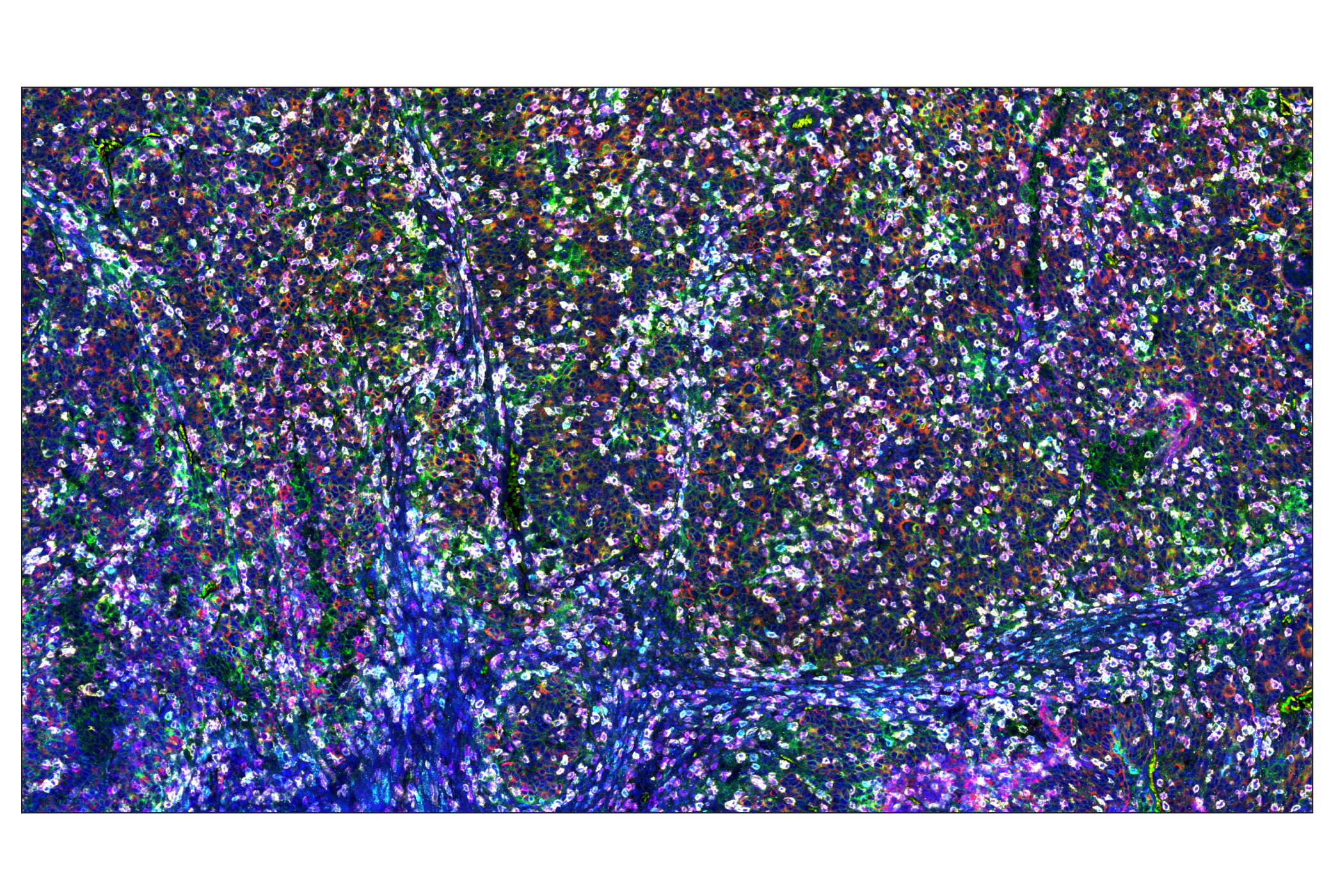
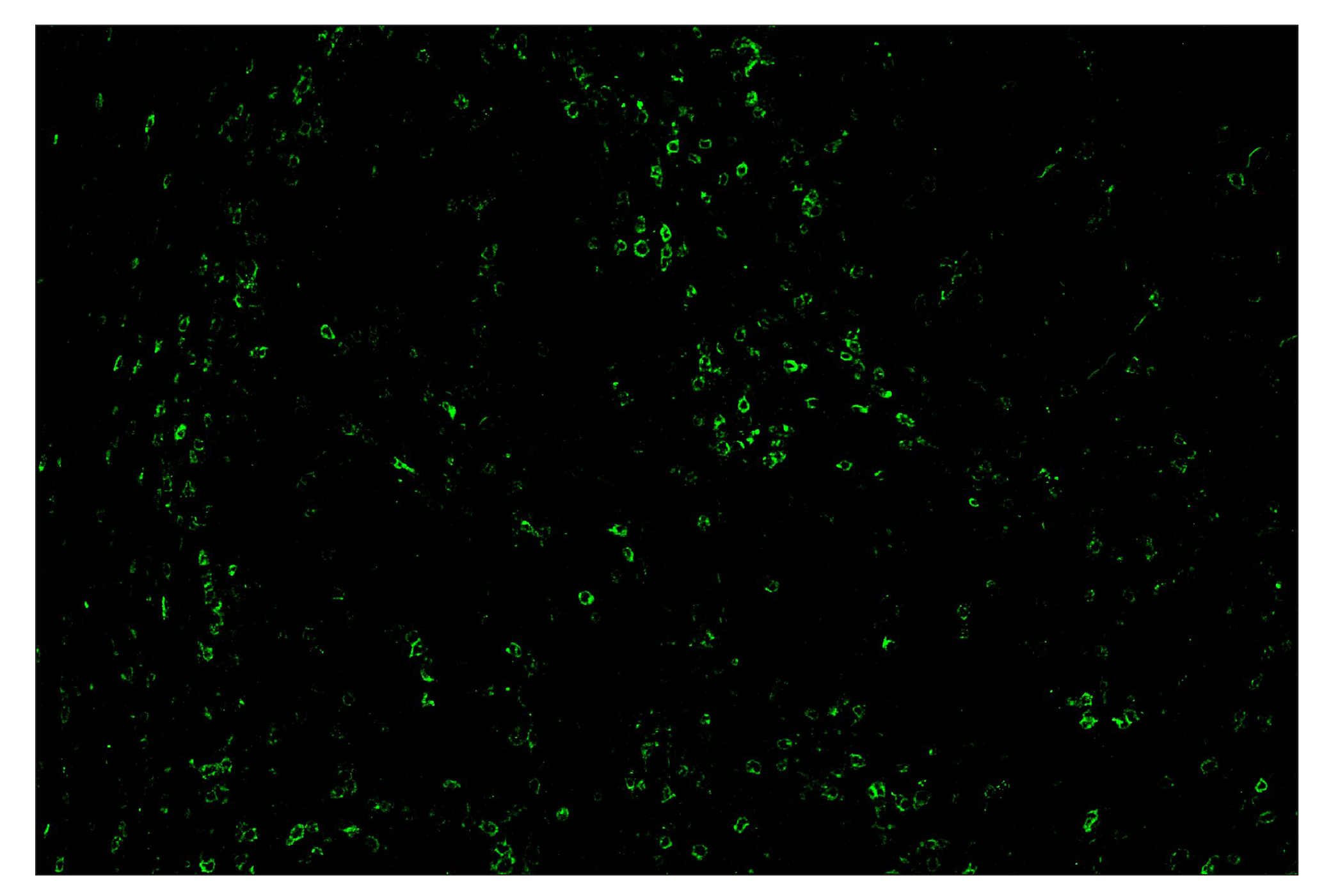
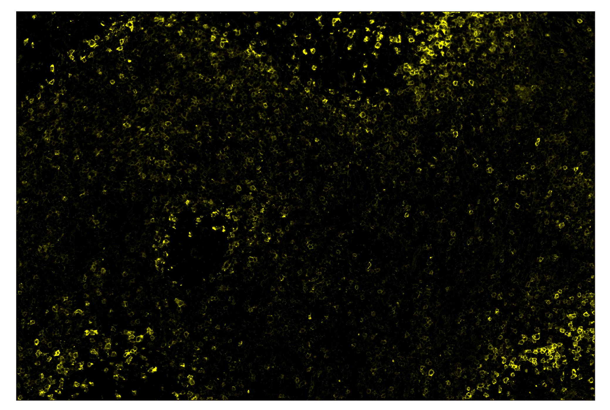
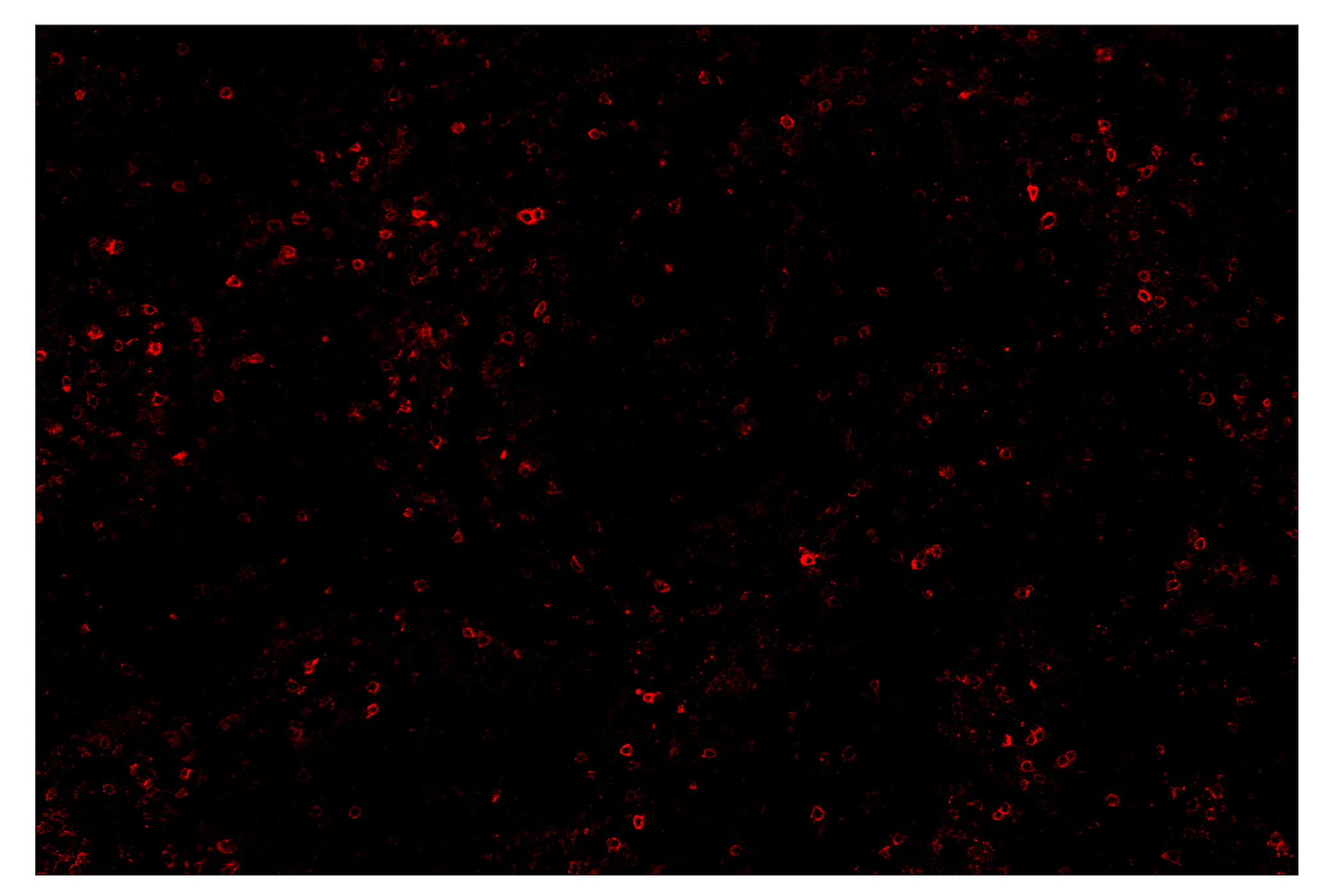
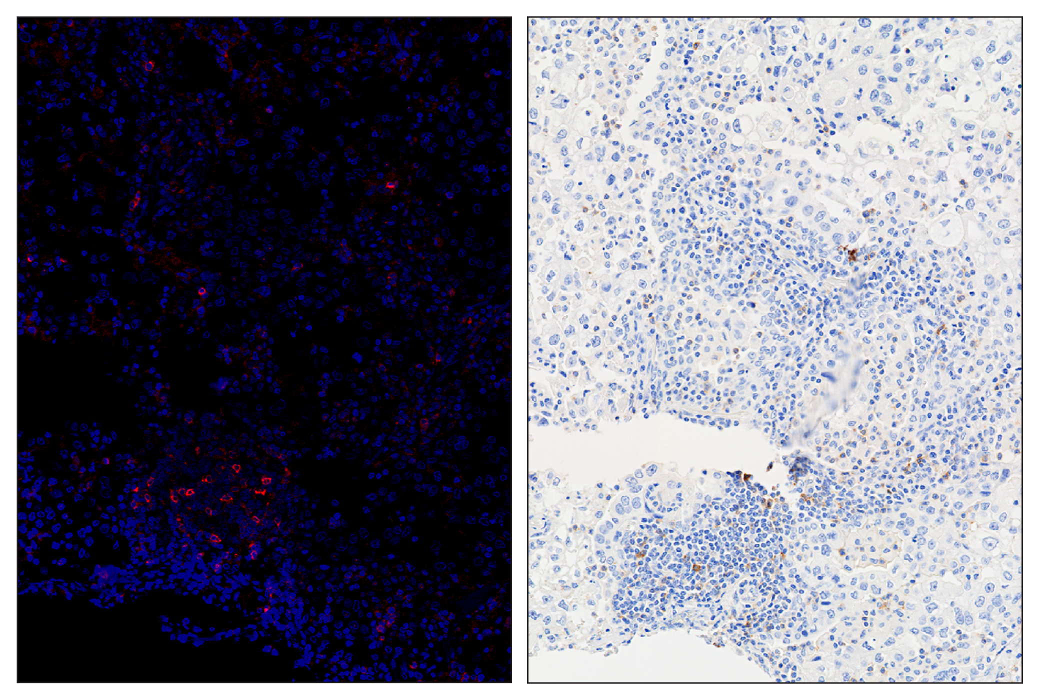
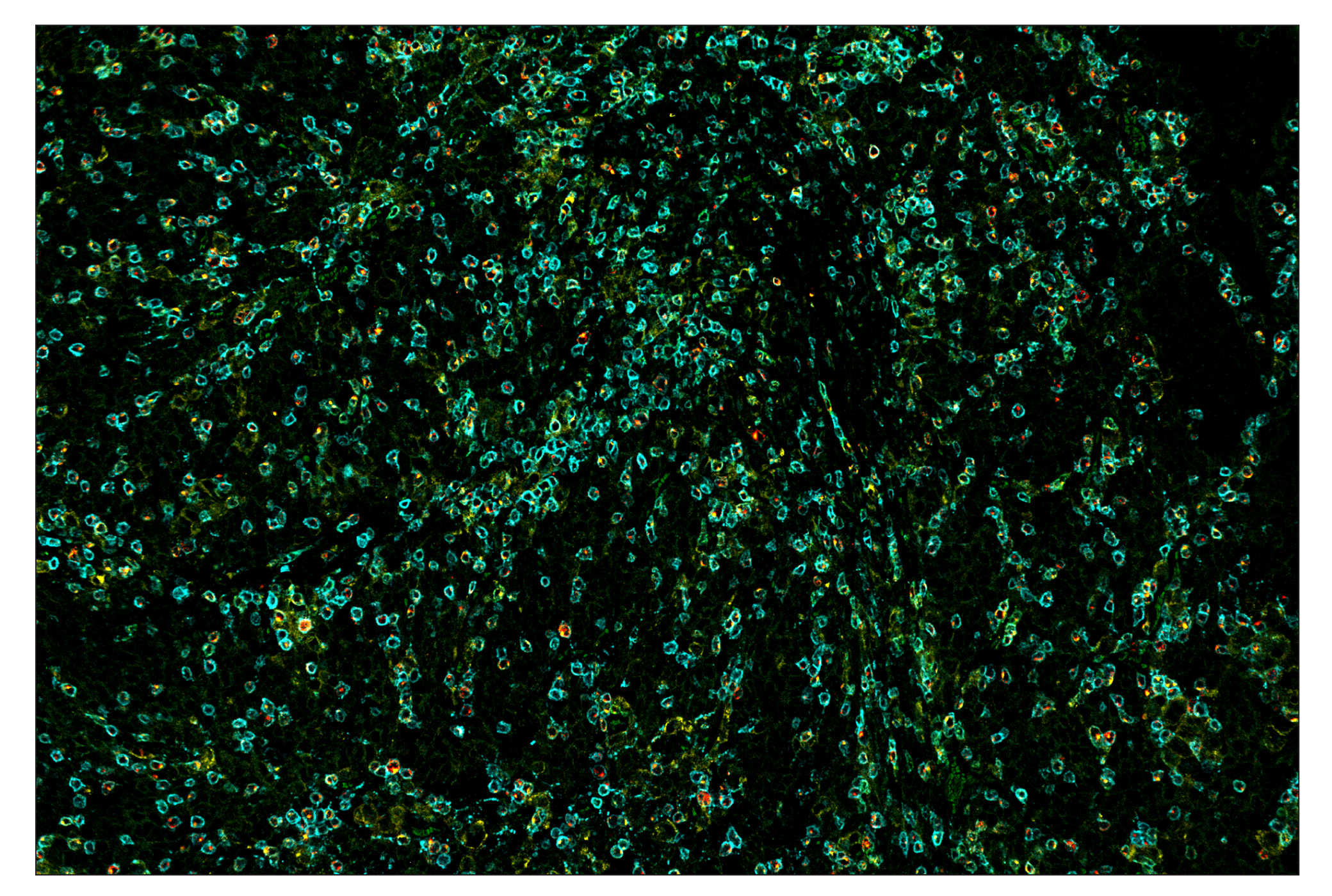
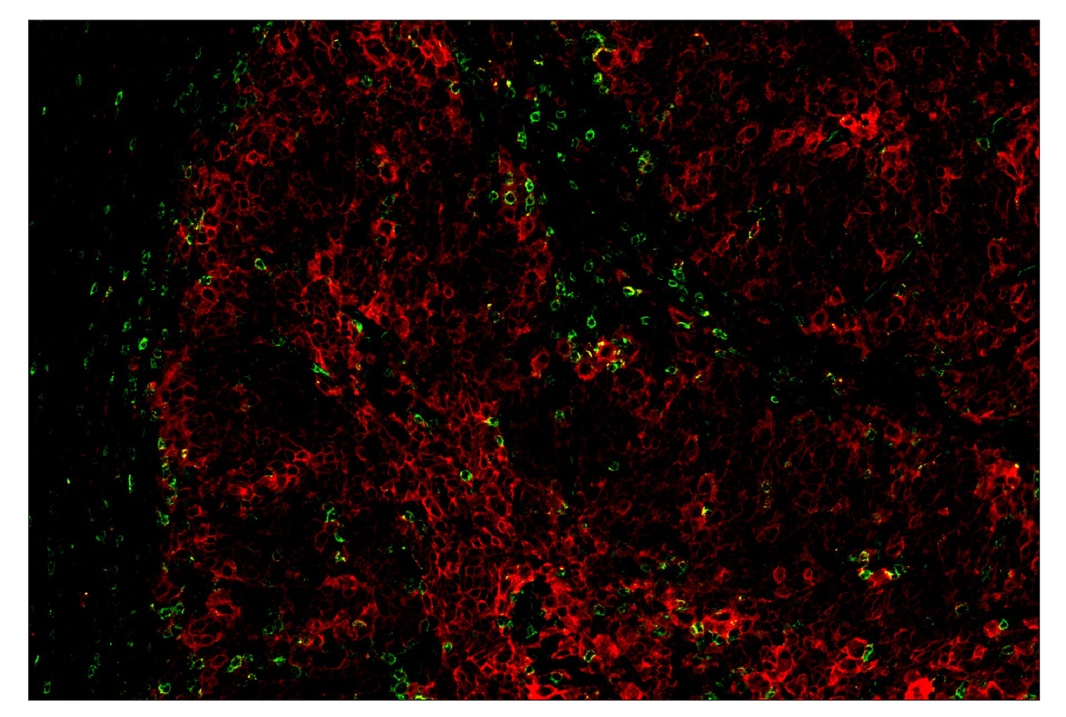
Monoclonal antibody is produced by immunizing animals with a synthetic peptide corresponding to residues surrounding Ala274 of human PD-1 protein.

Background

The programmed cell death 1 protein (PD-1, PDCD1, CD279) is a member of the CD28 family of immunoreceptors that regulate T cell activation and immune responses (1-3). The PD-1 protein contains an extracellular Ig V domain, a transmembrane domain, and a cytoplasmic tail that includes an immunoreceptor tyrosine-based inhibitory motif (ITIM) and an immunoreceptor tyrosine-based switch motif (ITSM). PD-1 is activated by the cell surface ligands PD-L1 and PD-L2 (4). Upon activation, PD-1 ITIM and ITSM phosphorylation leads to the recruitment of the protein tyrosine phosphatases SHP-1 and SHP-2, which suppress TCR signaling (5-7). In addition to activated T cells, PD-1 is expressed in activated B cells and monocytes, although its function in these cell types has not been fully characterized (8). The PD-1 pathway plays an important role in immune tolerance (3); however, research studies show that cancer cells often adopt this pathway to escape immune surveillance (9). Consequently, blockade of PD-1 and its ligands is proving to be a sound strategy for neoplastic intervention (10).

Limited Uses

Except as otherwise expressly agreed in a writing signed by a legally authorized representative of CST, the following terms apply to Products provided by CST, its affiliates or its distributors. Any Customer's terms and conditions that are in addition to, or different from, those contained herein, unless separately accepted in writing by a legally authorized representative of CST, are rejected and are of no force or effect.

Products are labeled with For Research Use Only or a similar labeling statement and have not been approved, cleared, or licensed by the FDA or other regulatory foreign or domestic entity, for any purpose. Customer shall not use any Product for any diagnostic or therapeutic purpose, or otherwise in any manner that conflicts with its labeling statement. Products sold or licensed by CST are provided for Customer as the end-user and solely for research and development uses. Any use of Product for diagnostic, prophylactic or therapeutic purposes, or any purchase of Product for resale (alone or as a component) or other commercial purpose, requires a separate license from CST. Customer shall (a) not sell, license, loan, donate or otherwise transfer or make available any Product to any third party, whether alone or in combination with other materials, or use the Products to manufacture any commercial products, (b) not copy, modify, reverse engineer, decompile, disassemble or otherwise attempt to discover the underlying structure or technology of the Products, or use the Products for the purpose of developing any products or services that would compete with CST products or services, (c) not alter or remove from the Products any trademarks, trade names, logos, patent or copyright notices or markings, (d) use the Products solely in accordance with CST Product Terms of Sale and any applicable documentation, and (e) comply with any license, terms of service or similar agreement with respect to any third party products or services used by Customer in connection with the Products.

For Research Use Only. Not for Use in Diagnostic Procedures.
Cell Signaling Technology is a trademark of Cell Signaling Technology, Inc.
SignalStar is a trademark of Cell Signaling Technology, Inc.
XP is a registered trademark of Cell Signaling Technology, Inc.
All other trademarks are the property of their respective owners. Visit our Trademark Information page.
To Purchase # 35347
Cat. # Size Qty. Price
35347S
1 Kit  (10 slides)